Your Ana pattern nuclear speckled images are ready in this website. Ana pattern nuclear speckled are a topic that is being searched for and liked by netizens today. You can Find and Download the Ana pattern nuclear speckled files here. Find and Download all royalty-free photos.
If you’re searching for ana pattern nuclear speckled pictures information linked to the ana pattern nuclear speckled interest, you have pay a visit to the right site. Our site frequently provides you with hints for viewing the highest quality video and picture content, please kindly hunt and locate more informative video articles and images that match your interests.
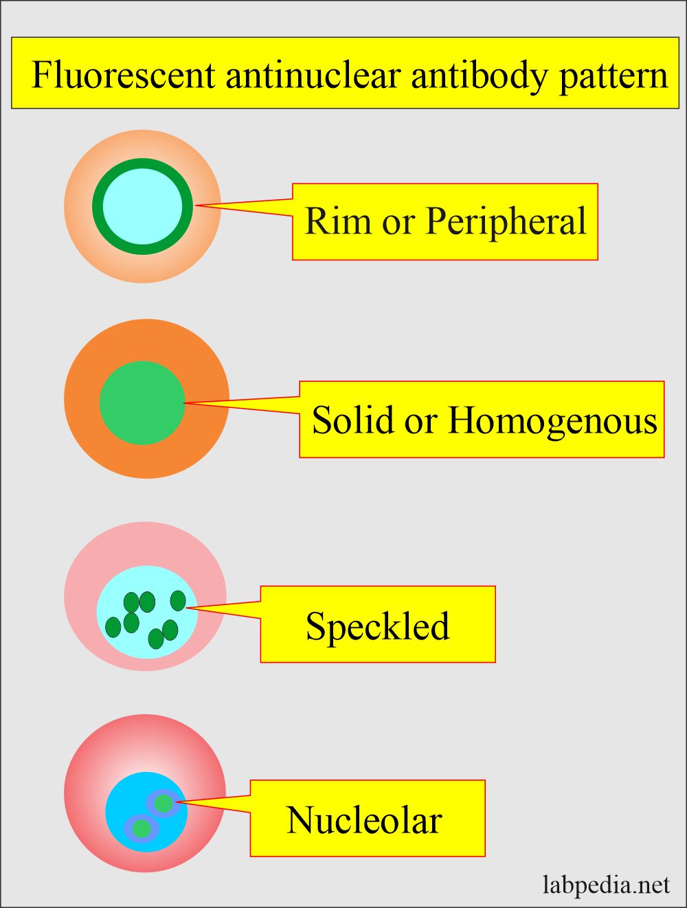
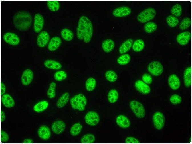
Ana Pattern Nuclear Speckled. 1d may not be reported depending on the. The presence of ANA with a homogeneous speckled HS pattern was significantly associated with the absence of cancer p 001. Speckled Dense fine speckled AC-2. A homogenous diffuse pattern appears as total nuclear fluorescence and is common in people with systemic lupus.
 Anti Dna Anti Double Stranded Dna Antibodies Anti Ds Dna Ab And Their Significance Labpedia Net From labpedia.net
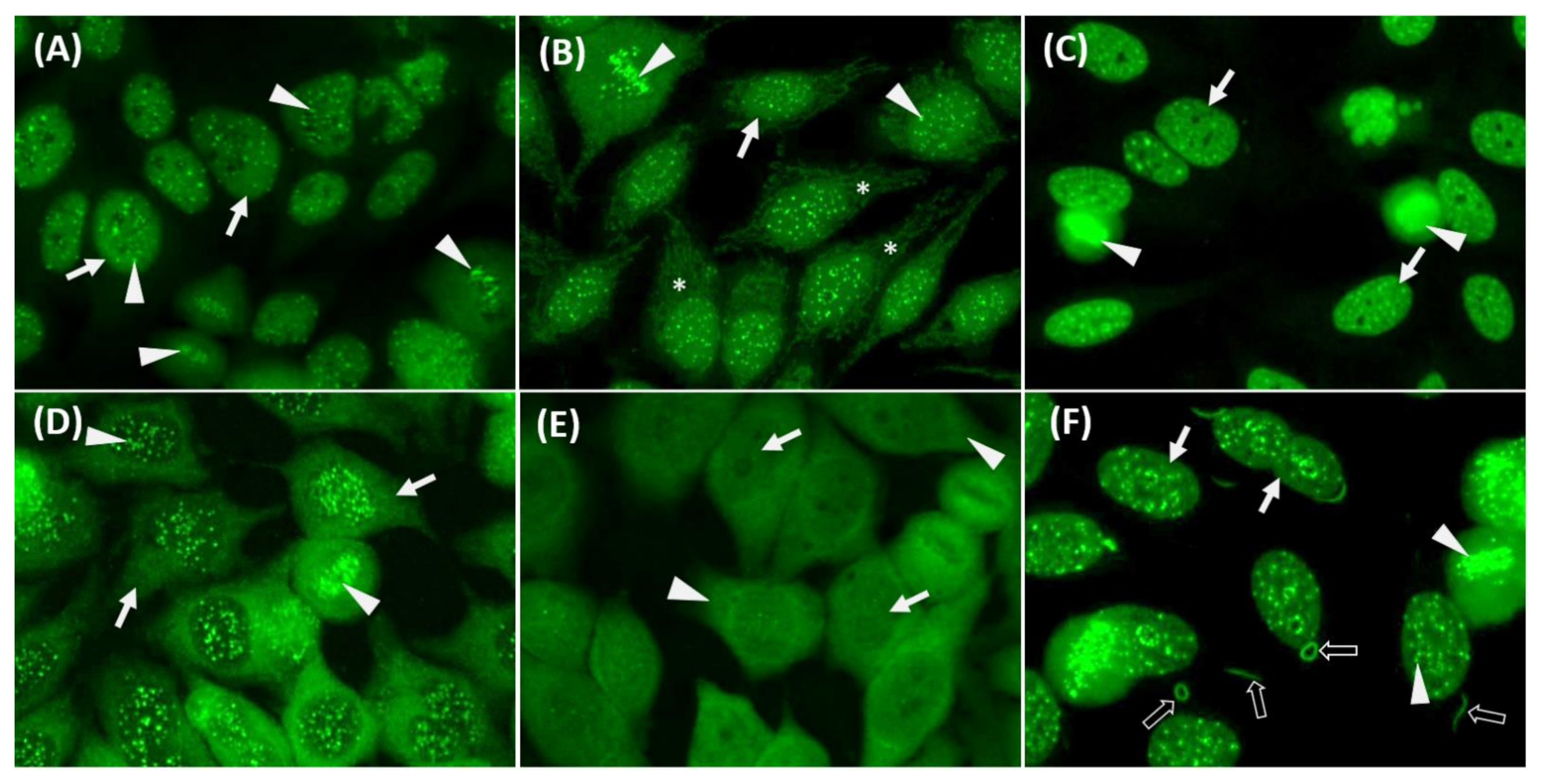
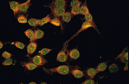
Speckled Dense fine speckled AC-2. Nuclear speckled pattern with striking variability in intensity with the strongest staining in G2 phase and weakestnegative staining in G1. It is found in many disease states including SLE and scleroderma. Some samples yield a nuclear speckled pattern with similar staining at the mitotic chromatin metaphase and anaphase very similar to AC-2 nuclear dense fine speckled pattern but do not yield a positive result in immunoassays specific for anti-DFS70 antibodies. The speckled pattern is commonly associated with lupus but is not enough to make a. A centromere pattern is usually reported as distinct pattern but they can also be termed discrete speckled nuclear staining patterns Fig.
A speckled pattern in an anti-nuclear antibodies test may indicate Sjogren syndrome scleroderma polymyositis rheumatoid arthritis or mixed connective tissue disease according to Lab Tests Online.
Nuclear speckled pattern with striking variability in intensity with the strongest staining in G2 phase and weakestnegative staining in G1. Cytoplasmic speckled patterns are a common finding and are associated with various antibodies including anti-synthetase antibodies. The pattern of the ANA test can give information about the type of autoimmune disease present and the appropriate treatment program. The speckled pattern is commonly associated with lupus but is not enough to make a. Prometaphase cells frequently show a weak staining of the nuclear envelope. ANA tests by indirect.
 Source: researchgate.net
The centromeres are positive only in prometaphase and metaphase revealing multiple aligned small and faint dots. Prometaphase cells frequently show a weak staining of the nuclear envelope. Speckled Dense fine speckled AC-2. It is found in many disease states including SLE and scleroderma. Speckled pattern correlates with antibody to nuclear antigens extractable by saline.
 Source: youtube.com
It is found in many disease states including SLE and scleroderma. An ANA of 1160 is a low titter and can be seen in healthy people. It is important to realize that even though 98 of people with lupus will have a positive ANA ANAs are also present in healthy individuals 5-10 and people with other connective tissue diseases such as scleroderma. Prometaphase cells frequently show a weak staining of the nuclear envelope. The patterns then can sometimes give the doctor further clues as to types of illnesses to look for in evaluating a patient.
 Source: labpedia.net
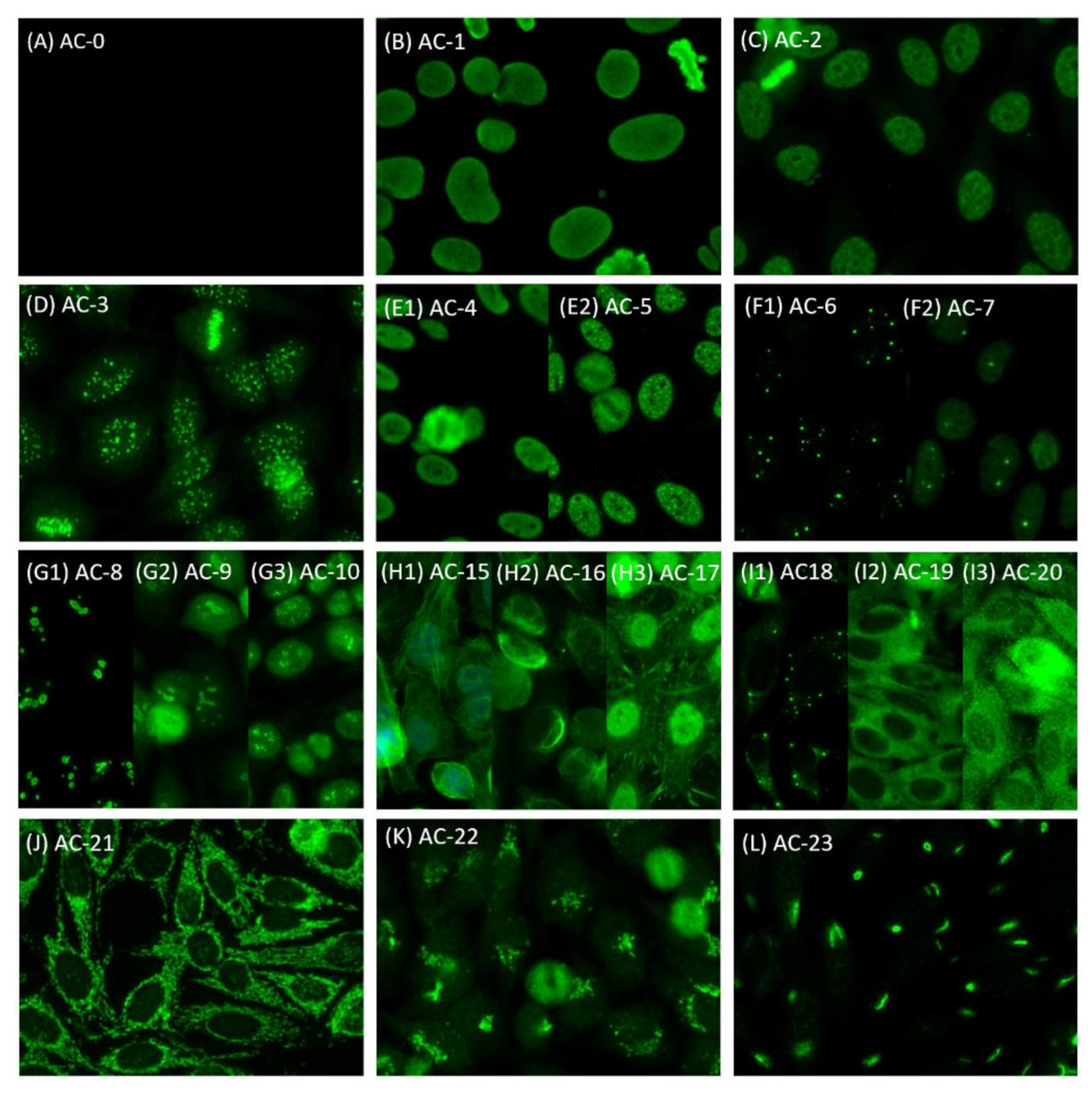
The pattern of the ANA test can give information about the type of autoimmune disease present and the appropriate treatment program. Major Group Major Pattern Groups Minor Pattern Subgroups AC Codes. Numerous small and uniform. One particular ANA pattern without a confirmed clinical correlation is the nuclear dense fine speckled ANA-DFS pattern. A uniform true speckled pattern may be s een with centromere antibodies in cells not in division.
 Source: mdpi.com
Immunofluorescence on HEp2-cells is the standard diagnostic assay for the detection of anti-nuclear antibodies ANA. The speckled pattern is seen in many conditions and in people who do not have any autoimmune disease. A centromere pattern is usually reported as distinct pattern but they can also be termed discrete speckled nuclear staining patterns Fig. Fine speckled pattern chromosome-negative. Anti-Scl70 and anti-RNAP-III were associated with cancer in 15 and 14 respectively.
 Source: researchgate.net
When antibodies to DNA and deoxyribonucleoprotein are present rim and homogenous pattern there may be interference with the detection of speckled pattern. A homogenous diffuse pattern appears as total nuclear fluorescence and is common in people with systemic lupus. The nuclear dense fine speckled DFS pattern is one of the most commonly observed IIF-ANA patterns in patients who are ANA-positive have no evident diagnosis of ANA-associated rheumatic autoimmune diseases AARD and have been referred to clinical laboratories for ANA testing because of non-specific complaints and symptoms 4-6. The group has defined six nuclear patterns as Competent-Level. Numerous small and uniform.
 Source: researchgate.net
These specific nuclear antibodies are themselves associated with specific autoimmune diseases. The pattern of the ANA test can give information about the type of autoimmune disease present and the appropriate treatment program. It is found in many disease states including SLE and scleroderma. Nuclear speckled pattern with striking variability in intensity with the strongest staining in G2 phase and weakestnegative staining in G1. One particular ANA pattern without a confirmed clinical correlation is the nuclear dense fine speckled ANA-DFS pattern.
 Source: researchgate.net
ANA tests by indirect. The presence of ANA with a homogeneous speckled HS pattern was significantly associated with the absence of cancer p 001. When antibodies to DNA and deoxyribonucleoprotein are present rim and homogenous pattern there may be interference with the detection of speckled pattern. Numerous small and uniform. 1d may not be reported depending on the.
 Source: mdpi.com
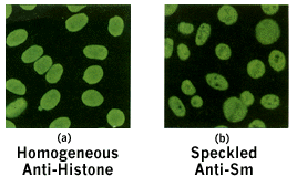
Speckled Patterns - The speckled pattern is the most commonly observed ANA pattern. Similarly a speckled pattern on the ANA test can indicate almost ANY autoimmune disease this is because a speckled pattern like a 180 titer level is not very significant. The patterns then can sometimes give the doctor further clues as to types of illnesses to look for in evaluating a patient. When antibodies to DNA and deoxyribonucleoprotein are present rim and homogenous pattern there may be interference with the detection of speckled pattern. ANA tests by indirect.
 Source: researchgate.net
Anti-DNA and anti-nuclear envelope antibodies cause this pattern. A homogenous diffuse pattern appears as total nuclear fluorescence and is common in people with systemic lupus. A speckled pattern may also appear on tests of individuals with systemic lupus states the Johns Hopkins Lupus Center. The patterns then can sometimes give the doctor further clues as to types of illnesses to look for in evaluating a patient. However classic ENA testing generally identifies only anti-Jo-1.
 Source: researchgate.net
When antibodies to DNA and deoxyribonucleoprotein are present rim and homogenous pattern there may be interference with the detection of speckled pattern. However classic ENA testing generally identifies only anti-Jo-1. When antibodies to DNA and deoxyribonucleoprotein are present rim and homogenous pattern there may be interference with the detection of speckled pattern. The speckled pattern is seen in many conditions and in people who do not have any autoimmune disease. A uniform true speckled pattern may be s een with centromere antibodies in cells not in division.
 Source: researchgate.net
Anti-DNA and anti-nuclear envelope antibodies cause this pattern. Each of these patterns possibly indicate the presence of specific nuclear antibodies. The centromeres are positive only in prometaphase and metaphase revealing multiple aligned small and faint dots. As defined by ICAP Chan et al 2015 the ANA-DFS pattern by IIF is characterized by a dense and heterogeneous speckled staining of both the nucleoplasm of interphase cells and the chromosomal plate of metaphase cells The ANA. It can be present in MCTD mixed connective tissue disorder Sjogrens syndrome lupus and many more autoimmune disease but the pattern is MOST commonly present in.
 Source: euroimmunblog.com
Prometaphase cells frequently show a weak staining of the nuclear envelope. It is found in many disease states including SLE and scleroderma. A speckled pattern may also appear on tests of individuals with systemic lupus states the Johns Hopkins Lupus Center. For example the nucleolar pattern is more commonly seen in the disease scleroderma. However classic ENA testing generally identifies only anti-Jo-1.
 Source: webpath.med.utah.edu
The patterns then can sometimes give the doctor further clues as to types of illnesses to look for in evaluating a patient. Dense fine speckled DFS pattern in antinuclear antibody ANA test using indirect immunofluorescence method became to be known recently and it is detected in patients with various chronic inflammatory diseases as well as in healthy individuals. Speckled Dense fine speckled AC-2. Fine speckled pattern chromosome-negative. The pattern of the ANA test can give information about the type of autoimmune disease present and the appropriate treatment program.
 Source: oooojournal.net
Each of these patterns possibly indicate the presence of specific nuclear antibodies. The speckled pattern is commonly associated with lupus but is not enough to make a. Major Group Major Pattern Groups Minor Pattern Subgroups AC Codes. The centromeres are positive only in prometaphase and metaphase revealing multiple aligned small and faint dots. The group has defined six nuclear patterns as Competent-Level.
 Source: news-medical.net
Cytoplasmic speckled patterns are a common finding and are associated with various antibodies including anti-synthetase antibodies. A centromere pattern is usually reported as distinct pattern but they can also be termed discrete speckled nuclear staining patterns Fig. ICAP recommends that any laboratory performing ANA by IIF should be able to accurately and reproducibly identify these patterns. Anti-Scl70 and anti-RNAP-III were associated with cancer in 15 and 14 respectively. It is important to realize that even though 98 of people with lupus will have a positive ANA ANAs are also present in healthy individuals 5-10 and people with other connective tissue diseases such as scleroderma.
 Source: cyberounds.com
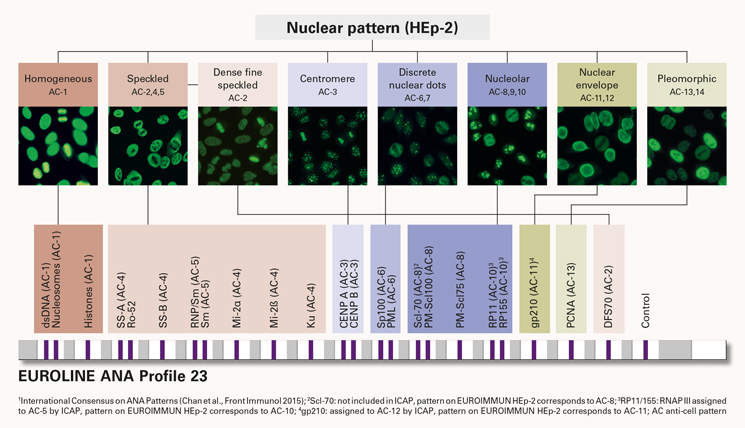
Discrete nuclear dots Centromere AC-3. Some samples yield a nuclear speckled pattern with similar staining at the mitotic chromatin metaphase and anaphase very similar to AC-2 nuclear dense fine speckled pattern but do not yield a positive result in immunoassays specific for anti-DFS70 antibodies. The pattern of the ANA test can give information about the type of autoimmune disease present and the appropriate treatment program. Another pattern known as a nucleolar pattern is common in people with scleroderma. As defined by ICAP Chan et al 2015 the ANA-DFS pattern by IIF is characterized by a dense and heterogeneous speckled staining of both the nucleoplasm of interphase cells and the chromosomal plate of metaphase cells The ANA.

The centromeres are positive only in prometaphase and metaphase revealing multiple aligned small and faint dots. Prometaphase cells frequently show a weak staining of the nuclear envelope. ICAP recommends that any laboratory performing ANA by IIF should be able to accurately and reproducibly identify these patterns. One particular ANA pattern without a confirmed clinical correlation is the nuclear dense fine speckled ANA-DFS pattern. It is important to realize that even though 98 of people with lupus will have a positive ANA ANAs are also present in healthy individuals 5-10 and people with other connective tissue diseases such as scleroderma.
 Source: researchgate.net
The nuclear dense fine speckled DFS pattern is one of the most commonly observed IIF-ANA patterns in patients who are ANA-positive have no evident diagnosis of ANA-associated rheumatic autoimmune diseases AARD and have been referred to clinical laboratories for ANA testing because of non-specific complaints and symptoms 4-6. Speckled Dense fine speckled AC-2. It is found in many disease states including SLE and scleroderma. For example the presence of a speckled positive ANA indicates the presence of these specific autoantibodies SSA SSB RNP Smith and Ku antibodies. Another pattern known as a nucleolar pattern is common in people with scleroderma.
This site is an open community for users to do submittion their favorite wallpapers on the internet, all images or pictures in this website are for personal wallpaper use only, it is stricly prohibited to use this wallpaper for commercial purposes, if you are the author and find this image is shared without your permission, please kindly raise a DMCA report to Us.
If you find this site helpful, please support us by sharing this posts to your own social media accounts like Facebook, Instagram and so on or you can also save this blog page with the title ana pattern nuclear speckled by using Ctrl + D for devices a laptop with a Windows operating system or Command + D for laptops with an Apple operating system. If you use a smartphone, you can also use the drawer menu of the browser you are using. Whether it’s a Windows, Mac, iOS or Android operating system, you will still be able to bookmark this website.


