Your Impaired relaxation pattern of lv diastolic filling images are ready in this website. Impaired relaxation pattern of lv diastolic filling are a topic that is being searched for and liked by netizens today. You can Find and Download the Impaired relaxation pattern of lv diastolic filling files here. Download all royalty-free photos and vectors.
If you’re searching for impaired relaxation pattern of lv diastolic filling pictures information connected with to the impaired relaxation pattern of lv diastolic filling topic, you have pay a visit to the right blog. Our website frequently gives you hints for seeking the highest quality video and picture content, please kindly surf and find more enlightening video articles and graphics that fit your interests.
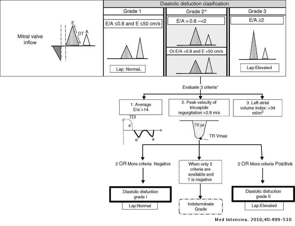
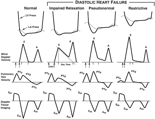
Impaired Relaxation Pattern Of Lv Diastolic Filling. Systolic failure presents reduced cardiac contractility whereas diastolic failure exhibits impaired cardiac relaxation with abnormal ventricular filling. Valsalva maneuver unmasks diastolic dysfunction and alters pseudnormal filling into impaired relaxation. In the presence of pseudonormal filling one will see a reversal of the pattern to that of impaired relaxation the EA ratio will drop below 1. Although this can be accomplished by radionuclide and computed tomography CT angiographic and.
 A Algorithm For Diagnosis Of Lv Diastolic Dysfunction In Subjects Download Scientific Diagram From researchgate.net
They increase cardiac output and decrease LV. Thus the Valsalva maneuver permits the investigator to unmask elevated filling pressures. In the presence of pseudonormal filling one will see a reversal of the pattern to that of impaired relaxation the EA ratio will drop below 1. Heart failure is responsible for a huge burden of disease in both developed and developing countries1 Among patients with heart failure about 50 show normal or preserved left ventricular LV systolic function HFpEF. The three basic events with respect to the left ventricle are LV contraction LV relaxation and LV filling. Although this can be accomplished by radionuclide and computed tomography CT angiographic and.
2 In the detection and evaluation of heart failure echocardiography plays a crucial role in evaluation of ventricular systolic function.
The three basic events with respect to the left ventricle are LV contraction LV relaxation and LV filling. In the presence of pseudonormal filling one will see a reversal of the pattern to that of impaired relaxation the EA ratio will drop below 1. The three basic events with respect to the left ventricle are LV contraction LV relaxation and LV filling. Although this can be accomplished by radionuclide and computed tomography CT angiographic and. Thus the Valsalva maneuver permits the investigator to unmask elevated filling pressures. Heart failure is responsible for a huge burden of disease in both developed and developing countries1 Among patients with heart failure about 50 show normal or preserved left ventricular LV systolic function HFpEF.
 Source: healio.com
2 In the detection and evaluation of heart failure echocardiography plays a crucial role in evaluation of ventricular systolic function. Valsalva maneuver unmasks diastolic dysfunction and alters pseudnormal filling into impaired relaxation. The three basic events with respect to the left ventricle are LV contraction LV relaxation and LV filling. Heart failure is responsible for a huge burden of disease in both developed and developing countries1 Among patients with heart failure about 50 show normal or preserved left ventricular LV systolic function HFpEF. It is calculated using Doppler echocardiography an ultrasound-based cardiac imaging modality.
 Source: researchgate.net
It is calculated using Doppler echocardiography an ultrasound-based cardiac imaging modality. Valsalva maneuver unmasks diastolic dysfunction and alters pseudnormal filling into impaired relaxation. Therefore at present assessing the type and degree of LV diastolic dysfunction relies on evaluating the pattern of LV filling. It is calculated using Doppler echocardiography an ultrasound-based cardiac imaging modality. Although this can be accomplished by radionuclide and computed tomography CT angiographic and.
 Source: br.pinterest.com
In the presence of pseudonormal filling one will see a reversal of the pattern to that of impaired relaxation the EA ratio will drop below 1. Thus the Valsalva maneuver permits the investigator to unmask elevated filling pressures. Therefore at present assessing the type and degree of LV diastolic dysfunction relies on evaluating the pattern of LV filling. Valsalva maneuver unmasks diastolic dysfunction and alters pseudnormal filling into impaired relaxation. It represents the ratio of peak velocity blood flow from left ventricular relaxation in early diastole the E wave to peak velocity flow in late diastole caused by atrial contraction the A wave.
 Source: medintensiva.org
Thus the Valsalva maneuver permits the investigator to unmask elevated filling pressures. 2 In the detection and evaluation of heart failure echocardiography plays a crucial role in evaluation of ventricular systolic function. Heart failure is responsible for a huge burden of disease in both developed and developing countries1 Among patients with heart failure about 50 show normal or preserved left ventricular LV systolic function HFpEF. They increase cardiac output and decrease LV. Systolic failure presents reduced cardiac contractility whereas diastolic failure exhibits impaired cardiac relaxation with abnormal ventricular filling.
 Source: researchgate.net
Although this can be accomplished by radionuclide and computed tomography CT angiographic and. Systolic failure presents reduced cardiac contractility whereas diastolic failure exhibits impaired cardiac relaxation with abnormal ventricular filling. Thus the Valsalva maneuver permits the investigator to unmask elevated filling pressures. Valsalva maneuver unmasks diastolic dysfunction and alters pseudnormal filling into impaired relaxation. In the presence of pseudonormal filling one will see a reversal of the pattern to that of impaired relaxation the EA ratio will drop below 1.
 Source: researchgate.net
2 In the detection and evaluation of heart failure echocardiography plays a crucial role in evaluation of ventricular systolic function. Although this can be accomplished by radionuclide and computed tomography CT angiographic and. Thus the Valsalva maneuver permits the investigator to unmask elevated filling pressures. It represents the ratio of peak velocity blood flow from left ventricular relaxation in early diastole the E wave to peak velocity flow in late diastole caused by atrial contraction the A wave. They increase cardiac output and decrease LV.
 Source: researchgate.net
They increase cardiac output and decrease LV. They increase cardiac output and decrease LV. It is calculated using Doppler echocardiography an ultrasound-based cardiac imaging modality. It represents the ratio of peak velocity blood flow from left ventricular relaxation in early diastole the E wave to peak velocity flow in late diastole caused by atrial contraction the A wave. Therefore at present assessing the type and degree of LV diastolic dysfunction relies on evaluating the pattern of LV filling.
 Source: cardioserv.net
Therefore at present assessing the type and degree of LV diastolic dysfunction relies on evaluating the pattern of LV filling. 2 In the detection and evaluation of heart failure echocardiography plays a crucial role in evaluation of ventricular systolic function. The EA ratio is a marker of the function of the left ventricle of the heart. The three basic events with respect to the left ventricle are LV contraction LV relaxation and LV filling. Reflecting the alternating pattern of hemorrhage and necrosis of zone 3 with the normal or slightly steatotic areas in zones 1 and 2.
 Source: researchgate.net
It represents the ratio of peak velocity blood flow from left ventricular relaxation in early diastole the E wave to peak velocity flow in late diastole caused by atrial contraction the A wave. The EA ratio is a marker of the function of the left ventricle of the heart. Reflecting the alternating pattern of hemorrhage and necrosis of zone 3 with the normal or slightly steatotic areas in zones 1 and 2. Thus the Valsalva maneuver permits the investigator to unmask elevated filling pressures. Systolic failure presents reduced cardiac contractility whereas diastolic failure exhibits impaired cardiac relaxation with abnormal ventricular filling.
 Source: 123sonography.com
Thus the Valsalva maneuver permits the investigator to unmask elevated filling pressures. Reflecting the alternating pattern of hemorrhage and necrosis of zone 3 with the normal or slightly steatotic areas in zones 1 and 2. They increase cardiac output and decrease LV. Valsalva maneuver unmasks diastolic dysfunction and alters pseudnormal filling into impaired relaxation. It is calculated using Doppler echocardiography an ultrasound-based cardiac imaging modality.
 Source: cardioserv.net
Valsalva maneuver unmasks diastolic dysfunction and alters pseudnormal filling into impaired relaxation. Valsalva maneuver unmasks diastolic dysfunction and alters pseudnormal filling into impaired relaxation. It represents the ratio of peak velocity blood flow from left ventricular relaxation in early diastole the E wave to peak velocity flow in late diastole caused by atrial contraction the A wave. Although this can be accomplished by radionuclide and computed tomography CT angiographic and. Systolic failure presents reduced cardiac contractility whereas diastolic failure exhibits impaired cardiac relaxation with abnormal ventricular filling.
 Source: researchgate.net
Valsalva maneuver unmasks diastolic dysfunction and alters pseudnormal filling into impaired relaxation. They increase cardiac output and decrease LV. Valsalva maneuver unmasks diastolic dysfunction and alters pseudnormal filling into impaired relaxation. Reflecting the alternating pattern of hemorrhage and necrosis of zone 3 with the normal or slightly steatotic areas in zones 1 and 2. Although this can be accomplished by radionuclide and computed tomography CT angiographic and.
 Source: criticalecho.com
Heart failure is responsible for a huge burden of disease in both developed and developing countries1 Among patients with heart failure about 50 show normal or preserved left ventricular LV systolic function HFpEF. Heart failure is responsible for a huge burden of disease in both developed and developing countries1 Among patients with heart failure about 50 show normal or preserved left ventricular LV systolic function HFpEF. They increase cardiac output and decrease LV. Although this can be accomplished by radionuclide and computed tomography CT angiographic and. Valsalva maneuver unmasks diastolic dysfunction and alters pseudnormal filling into impaired relaxation.
 Source: slidetodoc.com
2 In the detection and evaluation of heart failure echocardiography plays a crucial role in evaluation of ventricular systolic function. Although this can be accomplished by radionuclide and computed tomography CT angiographic and. It is calculated using Doppler echocardiography an ultrasound-based cardiac imaging modality. It represents the ratio of peak velocity blood flow from left ventricular relaxation in early diastole the E wave to peak velocity flow in late diastole caused by atrial contraction the A wave. The EA ratio is a marker of the function of the left ventricle of the heart.
 Source: researchgate.net
Although this can be accomplished by radionuclide and computed tomography CT angiographic and. Valsalva maneuver unmasks diastolic dysfunction and alters pseudnormal filling into impaired relaxation. 2 In the detection and evaluation of heart failure echocardiography plays a crucial role in evaluation of ventricular systolic function. It represents the ratio of peak velocity blood flow from left ventricular relaxation in early diastole the E wave to peak velocity flow in late diastole caused by atrial contraction the A wave. They increase cardiac output and decrease LV.
 Source: thoracickey.com
Systolic failure presents reduced cardiac contractility whereas diastolic failure exhibits impaired cardiac relaxation with abnormal ventricular filling. In the presence of pseudonormal filling one will see a reversal of the pattern to that of impaired relaxation the EA ratio will drop below 1. Therefore at present assessing the type and degree of LV diastolic dysfunction relies on evaluating the pattern of LV filling. Reflecting the alternating pattern of hemorrhage and necrosis of zone 3 with the normal or slightly steatotic areas in zones 1 and 2. Heart failure is responsible for a huge burden of disease in both developed and developing countries1 Among patients with heart failure about 50 show normal or preserved left ventricular LV systolic function HFpEF.
 Source: slidetodoc.com
Systolic failure presents reduced cardiac contractility whereas diastolic failure exhibits impaired cardiac relaxation with abnormal ventricular filling. Valsalva maneuver unmasks diastolic dysfunction and alters pseudnormal filling into impaired relaxation. Systolic failure presents reduced cardiac contractility whereas diastolic failure exhibits impaired cardiac relaxation with abnormal ventricular filling. Therefore at present assessing the type and degree of LV diastolic dysfunction relies on evaluating the pattern of LV filling. It is calculated using Doppler echocardiography an ultrasound-based cardiac imaging modality.
 Source: researchgate.net
Thus the Valsalva maneuver permits the investigator to unmask elevated filling pressures. 2 In the detection and evaluation of heart failure echocardiography plays a crucial role in evaluation of ventricular systolic function. Reflecting the alternating pattern of hemorrhage and necrosis of zone 3 with the normal or slightly steatotic areas in zones 1 and 2. It represents the ratio of peak velocity blood flow from left ventricular relaxation in early diastole the E wave to peak velocity flow in late diastole caused by atrial contraction the A wave. Valsalva maneuver unmasks diastolic dysfunction and alters pseudnormal filling into impaired relaxation.
This site is an open community for users to submit their favorite wallpapers on the internet, all images or pictures in this website are for personal wallpaper use only, it is stricly prohibited to use this wallpaper for commercial purposes, if you are the author and find this image is shared without your permission, please kindly raise a DMCA report to Us.
If you find this site value, please support us by sharing this posts to your favorite social media accounts like Facebook, Instagram and so on or you can also bookmark this blog page with the title impaired relaxation pattern of lv diastolic filling by using Ctrl + D for devices a laptop with a Windows operating system or Command + D for laptops with an Apple operating system. If you use a smartphone, you can also use the drawer menu of the browser you are using. Whether it’s a Windows, Mac, iOS or Android operating system, you will still be able to bookmark this website.


