Your The banding patterns of the dna fragments reveal that images are available in this site. The banding patterns of the dna fragments reveal that are a topic that is being searched for and liked by netizens today. You can Find and Download the The banding patterns of the dna fragments reveal that files here. Get all royalty-free photos.
If you’re searching for the banding patterns of the dna fragments reveal that images information linked to the the banding patterns of the dna fragments reveal that keyword, you have visit the right blog. Our site always provides you with hints for viewing the highest quality video and image content, please kindly search and locate more informative video content and images that match your interests.
The Banding Patterns Of The Dna Fragments Reveal That. In reality the logistics and technology used in such cases are rather straightforward. In order to identify a particular person with a high degree a certainty there must be a _____ probability that the DNA fingerprints from 2. Typically off-target DNA bands are caused by either partial digestion or Star Activity. Figure 173 Shown are DNA fragments from seven samples run on a gel stained with a fluorescent dye and viewed under UV light.
 Molecular Cloning Sequencing And Expression Of The Genes Encoding Adenosylcobalamin Dependent Diol Dehydrase Of Klebsiella Oxytoca Journal Of Biological Chemistry From jbc.org
Different banding patterns on a gel. However careful deployment of this technique and thoughtful interpretation of the fragment banding pattern can reveal the smallest differences in DNA sequences that account for the genetic variation among living things. Southern hybridization analysis revealed a high frequency of DNA polymorphism. Use of Multienzyme Multiplex PCR Amplified Fragment Length Polymorphism Typing in Analysis of Outbreaks of Multiresistant Klebsiella pneumoniae in an Intensive Care Unit Anneke van der Zee Niels Steer Eveline Thijssen Jolande Nelson Annemarie vant Veen and Anton Buiting. The DNA fragments shine up as bands. You need to compare your digestion to the expected DNA banding pattern.
If the DNA sequence differences that occur result in a longer DNA segment from one chromosome Fig.
The next step is to identify those bands to figure out which one to cut. The DNA fragments shine up as bands. Lane 1 contains the victims DNA fragments lane 2 the DNA evidence and lanes 3-6. If the bands in both lanes are similar to the expected pattern and the additional bands are limited to spaces within the upper and lower bands of the expected pattern the digestion is. DNA fingerprinting is a method for finding unique patterns in a persons DNA and using those patterns to identify people similar to how your unique fingerprint patterns can identify you. If the DNA sequence differences that occur result in a longer DNA segment from one chromosome Fig.
 Source: ncbi.nlm.nih.gov
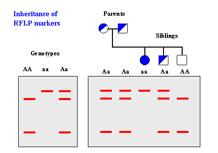
Heterozygous individuals will have two different versions of this DNA. Separation of DNA fragments on an agarose gel. Understanding how DNA banding patterns in a gel can aid in the conviction or exoneration of suspects and be utilized for positive identification of biological fathers in paternity cases can be intimidating. The same banding pattern just shows that these two samples are similiar in size. Restriction fragment length polymorphism RFLP is one of the easiest ways to study the diversity of the microbes.
 Source: numerade.com
So the phenotype is determined directly by the sequence of genetic material the genotype encodes. It binds to the DNA fragments in the gel. Agarose gel electrophoresis is an important technique in molecular genetics for a long. G-banding produces a banding pattern that can be correlated with Q-banding with the G-light bands equivalent to the Q-dull regions and the G-dark bands equivalent to the brightly fluorescent regions. Each band contains DNA fragments of the same size because they have travelled the same distance through the.
 Source: researchgate.net
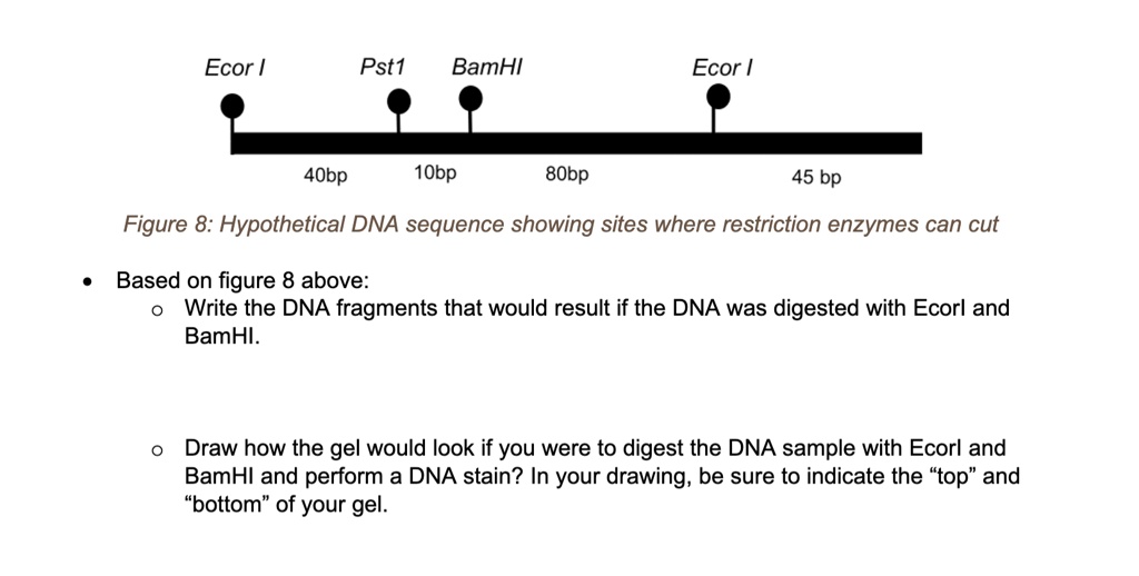
You need to compare your digestion to the expected DNA banding pattern. -the only way to determine if they are identical is by comparing and checking the two DNA sequences. A Complete Guide for Analysing and Interpreting Gel Electrophoresis Results. In the space below sketch the pattern of DNA fracments you would expect to see after performing an electrophoretic separation of a restriction dicest of a b and c above. Each band contains DNA fragments of the same size because they have travelled the same distance through the.
 Source: researchgate.net
If we know the sequence in which they cut then we know what the DNA sequence of the. Use of Multienzyme Multiplex PCR Amplified Fragment Length Polymorphism Typing in Analysis of Outbreaks of Multiresistant Klebsiella pneumoniae in an Intensive Care Unit Anneke van der Zee Niels Steer Eveline Thijssen Jolande Nelson Annemarie vant Veen and Anton Buiting. Species-specific patterns of DNA bending and sequence. Completely digested plasmid A. -the only way to determine if they are identical is by comparing and checking the two DNA sequences.
 Source: researchgate.net
G-banding produces a banding pattern that can be correlated with Q-banding with the G-light bands equivalent to the Q-dull regions and the G-dark bands equivalent to the brightly fluorescent regions. A chemical called ethidium bromide had been added to the gel. VanWye JD1 Bronson EC Anderson JN. Of the 97 strains analyzed 62 59 had the correct banding patterns detected by the bioanalyzer. In order to identify a particular person with a high degree a certainty there must be a _____ probability that the DNA fingerprints from 2.
 Source: ncbi.nlm.nih.gov
Typically off-target DNA bands are caused by either partial digestion or Star Activity. Separation of DNA fragments on an agarose gel. Of the 97 strains analyzed 62 59 had the correct banding patterns detected by the bioanalyzer. G-banding produces a banding pattern that can be correlated with Q-banding with the G-light bands equivalent to the Q-dull regions and the G-dark bands equivalent to the brightly fluorescent regions. DNA fingerprinting is a method for finding unique patterns in a persons DNA and using those patterns to identify people similar to how your unique fingerprint patterns can identify you.
 Source: mdpi.com
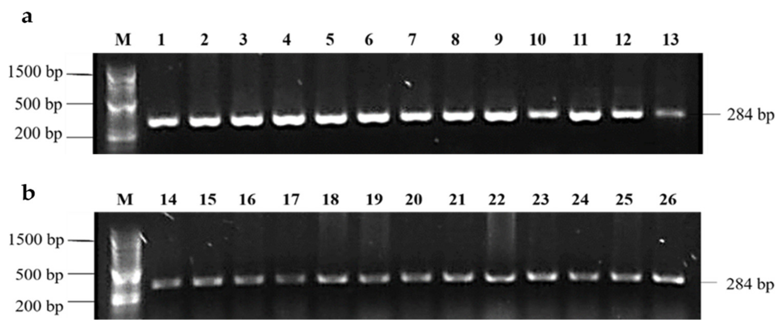
If the DNA sequence differences that occur result in a longer DNA segment from one chromosome Fig. Use of Multienzyme Multiplex PCR Amplified Fragment Length Polymorphism Typing in Analysis of Outbreaks of Multiresistant Klebsiella pneumoniae in an Intensive Care Unit Anneke van der Zee Niels Steer Eveline Thijssen Jolande Nelson Annemarie vant Veen and Anton Buiting. How DNA fingerprinting is made. RFLP patterns by conventional gel electrophoresis ranged from three to seven fragments for each strain. PCR Product with a faint primer dimer band.

It binds to the DNA fragments in the gel. A DNA fingerprint is a specific type of restriction map. Restriction fragment length polymorphism RFLP analysis compares DNA banding patterns of different DNA samples after restriction digestion Figure 1215. The cutinase gene probe did not reveal any polymorphisms. If the bands in both lanes are similar to the expected pattern and the additional bands are limited to spaces within the upper and lower bands of the expected pattern the digestion is.
 Source: bio.libretexts.org
RFLP has been successfully used for the study of diversity of V. The technique uses the simple restriction digestion of purified DNA from bacteria and variation in the banding pattern in the digestion reveals the genetic diversity. The cutinase gene probe did not reveal any polymorphisms. Each band contains DNA fragments of the same size because they have travelled the same distance through the. Of the 97 strains analyzed 62 59 had the correct banding patterns detected by the bioanalyzer.
 Source: courses.lumenlearning.com
It also fluoresces or lights up under UV light. Digested PCR product or DNA Fragment. The same banding pattern just shows that these two samples are similiar in size. PCR Product with a faint primer dimer band. As the DNA fragments move through the gel longer fragments are impeded more than shorter fragments producing characteristic banded patterns in the gel If cells from a carrot are removed and placed in a culture medium they can develop into a normal adult plant.
 Source: sciencedirect.com
If we know the sequence in which they cut then we know what the DNA sequence of the. There were 517 possible DNA fragments of which the bioanalyzer showed concordant results for 903 n 467 bands. -the only way to determine if they are identical is by comparing and checking the two DNA sequences. G-banding most commonly is introduced by treatment with a proteolytic enzyme such as trypsin followed by staining with Giemsa which binds DNA. The appearance of the banding pattern the size of the fragments tell us the DNA sequence of the plasmid because enzymes only cut at very specific sequences.
 Source: chegg.com
The same banding pattern just shows that these two samples are similiar in size. Agarose gel electrophoresis is an important technique in molecular genetics for a long. It shows _____ of DNA fragments in specific regions of a genome. This exercise is designed for use in high school environments as a stand. VanWye JD1 Bronson EC Anderson JN.
 Source: jbc.org
Southern hybridization analysis revealed a high frequency of DNA polymorphism. G-banding produces a banding pattern that can be correlated with Q-banding with the G-light bands equivalent to the Q-dull regions and the G-dark bands equivalent to the brightly fluorescent regions. How DNA fingerprinting is made. Of the 97 strains analyzed 62 59 had the correct banding patterns detected by the bioanalyzer. A mixture of genomic DNA fragments of varying sizes appear as a long smear whereas uncut genomic DNA is usually too large to run through the gel and forms a single large band at the top of the gel.
 Source: passel-old.unl.edu
The 6 lanes of DNA fragments exhibit banding patterns that can be used comparatively in this case to match evidence with a suspect. The technique uses the simple restriction digestion of purified DNA from bacteria and variation in the banding pattern in the digestion reveals the genetic diversity. Restriction fragment length polymorphism RFLP analysis compares DNA banding patterns of different DNA samples after restriction digestion Figure 1215. It binds to the DNA fragments in the gel. Restriction fragment length polymorphism RFLP is one of the easiest ways to study the diversity of the microbes.
 Source: nature.com
If we know the sequence in which they cut then we know what the DNA sequence of the. 1Department of Biological Sciences Purdue University West Lafayette IN 47907. Restriction fragments generated by each enzyme-probe combination resulted in distinct banding patterns clearly separating the isolates into two groups. Heterozygous individuals will have two different versions of this DNA. DNA bands can only be visualized using agarose gel electrophoresis.
 Source: courses.lumenlearning.com
The technique uses the simple restriction digestion of purified DNA from bacteria and variation in the banding pattern in the digestion reveals the genetic diversity. As the DNA fragments move through the gel longer fragments are impeded more than shorter fragments producing characteristic banded patterns in the gel If cells from a carrot are removed and placed in a culture medium they can develop into a normal adult plant. Completely digested plasmid A. In the space below sketch the pattern of DNA fracments you would expect to see after performing an electrophoretic separation of a restriction dicest of a b and c above. Species-specific patterns of DNA bending and sequence.
 Source: researchgate.net
Different banding patterns on a gel. However they may or may not have the same nucleotide sequence. Each band contains DNA fragments of the same size because they have travelled the same distance through the. Heterozygous individuals will have two different versions of this DNA. 1Department of Biological Sciences Purdue University West Lafayette IN 47907.
 Source: courses.lumenlearning.com
VanWye JD1 Bronson EC Anderson JN. It shows _____ of DNA fragments in specific regions of a genome. Thirty-one bands were not. Typically off-target DNA bands are caused by either partial digestion or Star Activity. In the space below sketch the pattern of DNA fracments you would expect to see after performing an electrophoretic separation of a restriction dicest of a b and c above.
This site is an open community for users to do sharing their favorite wallpapers on the internet, all images or pictures in this website are for personal wallpaper use only, it is stricly prohibited to use this wallpaper for commercial purposes, if you are the author and find this image is shared without your permission, please kindly raise a DMCA report to Us.
If you find this site helpful, please support us by sharing this posts to your own social media accounts like Facebook, Instagram and so on or you can also save this blog page with the title the banding patterns of the dna fragments reveal that by using Ctrl + D for devices a laptop with a Windows operating system or Command + D for laptops with an Apple operating system. If you use a smartphone, you can also use the drawer menu of the browser you are using. Whether it’s a Windows, Mac, iOS or Android operating system, you will still be able to bookmark this website.


